

Echographie pulmonaire et COVID-19 - Service de radiodiagnostic et radiologie interventionnelle - CHUV. Multiorgan POCUS in COVID-19: An Outpatient-Inpatient Approach - POCUS 101. Skip to content Let's Connect!
Twitter Youtube Instagram Facebook-f Linkedin Contact: Editor@POCUS101.com Menu Start typing and press enter to search Get Free POCUS Updates! Don't miss out. You have Successfully Subscribed! Pin It on Pinterest Share This. A simplified lung ultrasound for the diagnosis of interstitial lung disease in connective tissue disease: a meta-analysis. Point‐of‐care lung ultrasound in patients with COVID‐19: a narrative review. #POCUSforCOVID. Lung Ultrasound in COVID-19. Complete GUIDE to Lung Ultrasound in COVID-19 (Coronavirus) Patients - POCUS 101. Skip to content Connect With Us!
Youtube Twitter Instagram Facebook-f Contact: Editor@POCUS101.com © 2020 - Physician Zen LLC Menu Start typing and press enter to search Get Free POCUS Updates! Don't miss out. You have Successfully Subscribed! Pin It on Pinterest Share This. Proposal for international standardization of the use of lung ultrasound for COVID‐19 patients; a simple, quantitative, reproducible method - Soldati - - Journal of Ultrasound in Medicine. Covid-19 – Sonographic Tendencies. Given the pandemic status and constant coverage along with countries closing down, it is certain most of you have heard of Covid-19.

As healthcare workers including sonographers, radiographers, physicians, nurses, respiratory therapists, phlebotomists etc… We may come in contact or be affected by this pathogen. So what is Covid-19? Coronavirus disease 2019 (COVID-19) is an infectious disease caused by the severe acute respiratory syndrome coronavirus 2 (SARS-CoV-2). It is a novel coronavirus which was first detected in Wuhan, China. It has since spread to 178 countries and territories with the WHO declaring a pandemic on March, 11 2020. The virus is a zoonotic pathogen which spreads from animals to human. Symptoms Infection ranges from asymptomatic to severe. Covid-19 #POCUS resources - Zedu. Covid-19 #POCUS resources 18th Mar 20 A resource of the best in #POCUS ultrasound resources from the best in #POCUS providers around the world - dedicated to the fight against Covid19 Thank you to everyone who has poured their passion, time and energy into each and every one of these resources.

For our statement on how we as a small business are managing this dynamic and changing situation please click the button below. Covid-19: Lets stand together | The future of Zedu... This page is being updated daily, but the resources are no longer all displaying due to the sheer number! We have now separated and categorised the resources which can be accessed here. VIDEO: Imaging COVID-19 With Point-of-Care Ultrasound (POCUS) Regina Druz, M.D., FASNC, a member of the American Society of Nuclear Cardiology (ASNC) Board of Directors, chairwomen of the American College of Cardiology (ACC) Healthcare Innovation Section, and a cardiologist at Integrative Cardiology Center of Long Island, N.Y., explains the rapid expansion of telemedicine with the U.S. spread of novel coronavirus (COVID-19, SARS-CoV-2).
Druz spoken on the unprecedented expansion of telemedicine in the U.S., which more use in the last two weeks since states started calling for shelter in place orders, as opposed to the past two decades. The Centers for Medicare and Medicaid Services (CMS) previously only reimbursed for Telehealth in rural areas it determined had a shortage of doctors. However, in early March 2020, CMS dropped the geographic requirements and allowed Telehealth usage across th country as a way to mitigate person-to-person contact and keep vulnerable, older patients at home for routine check ups with doctors. APECHO article on line Anesthesiology 2020. 5 Minute Sono – Viral PNA – Core Ultrasound.
COVID and Machine Decontamination – Core Ultrasound. How lung ultrasound is helping clinicians with early diagnosis of Novel Coronavirus (COVID-19) Pneumonia - Gulfcoast Ultrasound News Blog. Written by: Trisha Reo, RDMS, RVT Bedside lung ultrasound is being recommended for all patients who present to the ED with flu-like symptoms for the early diagnosis and management of COVID-19 pneumonia.
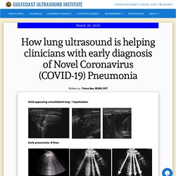
Lung ultrasound is highly sensitive and specific and considered as an alternative to chest radiography or CT scanning. It is performed in a matter of minutes and can be done at the bedside, minimizing the need to move the patient to various areas of the ED, therefore reducing the risk of exposure and spread of this highly infectious virus. As pneumonia progresses through stages, the appearance on ultrasound will vary depending on the degree and extent of consolidation. Fluid filled alveoli surrounded by air-filled lung creates short path reverberation artifact, which we call B-lines. As the pneumonia progresses to the next stage, purulent fluid fills the alveoli and the lung takes on a more solid appearance on ultrasound, similar to the appearance of the liver. Upcoming Webinars. Italian Doctor Triages by Ultrasound.
Editor's note: Find the latest COVID-19 news and guidance in Medscape's Coronavirus Resource Center .
Patients have been arriving at the emergency department of the University Hospital San Luigi Gonzaga, in Turin, Italy, one after another after another. At first, physicians thought advanced age was a good predictor of which cases might go downhill fast. They were wrong. "We intubated a woman who was 38 years old," says Giovanni Volpicelli, MD. Error - Cookies Turned Off. Findings of lung ultrasonography of novel corona virus pneumonia during the 2019–2020 epidemic. Findings of lung ultrasonography of novel corona virus pneumonia during the 2019–2020 epidemic. We performed lung ultrasonography on 20 patients with COVID-19 using a 12-zone method [3].
Characteristic findings included the following: 1.Thickening of the pleural line with pleural line irregularity;2.B lines in a variety of patterns including focal, multifocal, and confluent;3.Consolidations in a variety of patterns including multifocal small, non-translobar, and translobar with occasional mobile air bronchograms;4.Appearance of A lines during recovery phase;5.Pleural effusions are uncommon. The observed patterns occurred across a continuum from mild alveolar interstitial pattern, to severe bilateral interstitial pattern, to lung consolidation.
The role of lung ultrasound in the diagnosis of interstitial lung disease - Falcetta - Shanghai Chest. What is interstitial lung disease (ILD)?
ILD is defined as thickening of the pulmonary interstitium (space between the capillary endothelium and the alveolar epithelium) leading to impaired gas exchange due to various causes. ILD may be idiopathic or caused by exposure to organic and inorganic substances (i.e., hypersensitivity pneumonitis and pneumoconiosis), medical conditions [i.e., connective tissue diseases (CTDs), multisystemic diseases and obstructive sleep apnea], drugs, infection and radiation therapy (1,2). The overall estimated prevalence of ILD is about 25–74/100,000 population and up to 80.9 per 100,000 in men and 67.2 per 100,000 in women (3).
Diagnosis of ILD is usually made based on combination of clinical, functional, radiological and histological data. Chest X-ray (CXR) is often the first imaging test performed in ILD and British Thoracic Society recommend for high-resolution computed tomography (HRCT) if diagnosis is uncertain after CXR and clinical assessment. None. Should the ultrasound probe replace your stethoscope? A SICS-I sub-study comparing lung ultrasound and pulmonary auscultation in the critically ill. In this prospective observational study, we found poor agreement between auscultation and LUS for the diagnosis of pulmonary edema in acutely admitted critically ill patients.
Several previous studies focused on the diagnostic accuracy of LUS compared to other imaging modalities, such as chest X-ray and CT scan [4, 10, 20]. However, few studies have compared the diagnostic accuracy of LUS with the stethoscope, one of the most frequently used instruments at the bedside. Ultrasound GEL. 》Make sure you check out the Ultrasound Podcast COVID-19 episode for more discussion of how to use POCUS in this disease! 《 Background. ULA_covid - Free Lung Module Channel. Combatting COVID19 - Is Lung Ultrasound an Option? Written by Dr Cian McDermott, Emergency Physician & Director of Emergency Ultrasound Education, Mater University Hospital, Dublin, Ireland | @cianmcdermott Reviewed by Dr Rachel Liu, Emergency Physician & Director of Point-of-Care Ultrasound Education, Yale School of Medicine, New Haven, CT USA | @rubbleEM Whether you are the ultrasound educational lead for your hospital/ trust, a routine user or interested novice we’re sure that you will have seen lots of exciting data on twitter (using the #POCUSforCOVID hashtag), suggesting that USS has a role in the diagnosis and perhaps prognosis of Covid-19.
Maybe you’re self isolating right now and looking to get trained up on this new disease ready to return to the hospital soon. Like you, we were excited to read how Lung ultrasound (LUS) can show early changes that may be as reliable as CT and probably better than CXR. Covid-19 #POCUS resources - Zedu. COVID-19 Lung Module - Intelligent Ultrasound. COVID-19. Fighting COVID-19 together - Butterfly Network.