

Electroencephalography Simultaneous video and EEG recording of two guitarists improvising. Electroencephalography (EEG) is the recording of electrical activity along the scalp. EEG measures voltage fluctuations resulting from ionic current flows within the neurons of the brain.[1] In clinical contexts, EEG refers to the recording of the brain's spontaneous electrical activity over a short period of time, usually 20–40 minutes, as recorded from multiple electrodes placed on the scalp. Diagnostic applications generally focus on the spectral content of EEG, that is, the type of neural oscillations that can be observed in EEG signals. EEG is most often used to diagnose epilepsy, which causes obvious abnormalities in EEG readings.[2] It is also used to diagnose sleep disorders, coma, encephalopathies, and brain death. History[edit] Hans Berger In 1934, Fisher and Lowenback first demonstrated epileptiform spikes. In 1947, The American EEG Society was founded and the first International EEG congress was held.
Will Potter: The secret US prisons you've never heard of before | TED Talk Subtitles and Transcript Father Daniel Berrigan once said that "writing about prisonersis a little like writing about the dead."I think what he meant is that we treat prisoners as ghosts.They're unseen and unheard.It's easy to simply ignore themand it's even easier when the government goes to great lengths to keep them hidden. As a journalist, I think these storiesof what people in power do when no one is watching,are precisely the stories that we need to tell.That's why I began investigatingthe most secretive and experimental prison units in the United States,for so-called "second-tier" terrorists.The government calls these units Communications Management Units or CMUs.Prisoners and guards call them "Little Guantanamo."They are islands unto themselves.But unlike Gitmo they exist right here, at home,floating within larger federal prisons. There's an estimated 60 to 70 prisoners here,and they're overwhelmingly Muslim.They include people like Dr. So, why was he moved? (Laughter) For the record, I'm not. Thank you.
Psilocybin, the Drug in Magic Mushrooms, Lifts Mood and Increases Compassion Over the Long Term - - TIME Healthland - StumbleUpon The psychedelic drug in magic mushrooms may have lasting medical and spiritual benefits, according to new research from Johns Hopkins School of Medicine. The mushroom-derived hallucinogen, called psilocybin, is known to trigger transformative spiritual states, but at high doses it can also result in “bad trips” marked by terror and panic. The trick is to get the dose just right, which the Johns Hopkins researchers report having accomplished. In their study, the Hopkins scientists were able to reliably induce transcendental experiences in volunteers, which offered long-lasting psychological growth and helped people find peace in their lives — without the negative effects. (PHOTOS: Inside Colorado’s Marijuana Industry) “The important point here is that we found the sweet spot where we can optimize the positive persistent effects and avoid some of the fear and anxiety that can occur and can be quite disruptive,” says lead author Roland Griffiths, professor of behavioral biology at Hopkins.
Brain Atlas - Introduction The central nervous system (CNS) consists of the brain and the spinal cord, immersed in the cerebrospinal fluid (CSF). Weighing about 3 pounds (1.4 kilograms), the brain consists of three main structures: the cerebrum, the cerebellum and the brainstem. Cerebrum - divided into two hemispheres (left and right), each consists of four lobes (frontal, parietal, occipital and temporal). – closely packed neuron cell bodies form the grey matter of the brain. Cerebellum – responsible for psychomotor function, the cerebellum co-ordinates sensory input from the inner ear and the muscles to provide accurate control of position and movement. Brainstem – found at the base of the brain, it forms the link between the cerebral cortex, white matter and the spinal cord. Other important areas in the brain include the basal ganglia, thalamus, hypothalamus, ventricles, limbic system, and the reticular activating system. Basal Ganglia Thalamus and Hypothalamus Ventricles Limbic System Reticular Activating System Glia
Diffusion MRI Diffusion MRI (or dMRI) is a magnetic resonance imaging (MRI) method which came into existence in the mid-1980s.[1][2][3] It allows the mapping of the diffusion process of molecules, mainly water, in biological tissues, in vivo and non-invasively. Molecular diffusion in tissues is not free, but reflects interactions with many obstacles, such as macromolecules, fibers, membranes, etc. Water molecule diffusion patterns can therefore reveal microscopic details about tissue architecture, either normal or in a diseased state. The first diffusion MRI images of the normal and diseased brain were made public in 1985.[4][5] Since then, diffusion MRI, also referred to as diffusion tensor imaging or DTI (see section below) has been extraordinarily successful. In diffusion weighted imaging (DWI), the intensity of each image element (voxel) reflects the best estimate of the rate of water diffusion at that location. Diffusion[edit] Given the concentration and flux where D is the diffusion coefficient.
Functional magnetic resonance imaging Researcher checking fMRI images Functional magnetic resonance imaging or functional MRI (fMRI) is a functional neuroimaging procedure using MRI technology that measures brain activity by detecting associated changes in blood flow.[1] This technique relies on the fact that cerebral blood flow and neuronal activation are coupled. When an area of the brain is in use, blood flow to that region also increases. The primary form of fMRI uses the Blood-oxygen-level dependent (BOLD) contrast,[2] discovered by Seiji Ogawa. The procedure is similar to MRI but uses the change in magnetization between oxygen-rich and oxygen-poor blood as its basic measure. FMRI is used both in the research world, and to a lesser extent, in the clinical world. Overview[edit] The fMRI concept builds on the earlier MRI scanning technology and the discovery of properties of oxygen-rich blood. History[edit] Three studies in 1992 were the first to explore using the BOLD contrast in humans. Physiology[edit]
Neuron All neurons are electrically excitable, maintaining voltage gradients across their membranes by means of metabolically driven ion pumps, which combine with ion channels embedded in the membrane to generate intracellular-versus-extracellular concentration differences of ions such as sodium, potassium, chloride, and calcium. Changes in the cross-membrane voltage can alter the function of voltage-dependent ion channels. If the voltage changes by a large enough amount, an all-or-none electrochemical pulse called an action potential is generated, which travels rapidly along the cell's axon, and activates synaptic connections with other cells when it arrives. Neurons do not undergo cell division. In most cases, neurons are generated by special types of stem cells. A type of glial cell, called astrocytes (named for being somewhat star-shaped), have also been observed to turn into neurons by virtue of the stem cell characteristic pluripotency. Overview[edit] Anatomy and histology[edit]
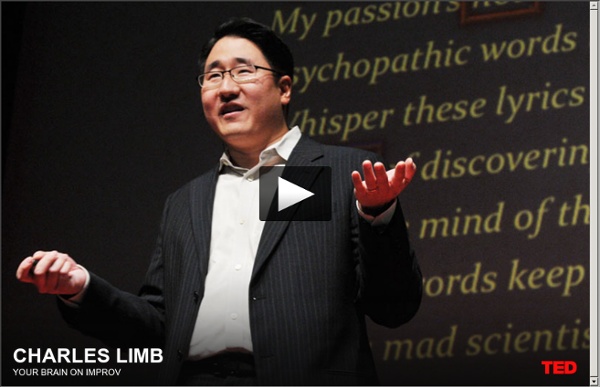
Charles Limb studies the neurology of music and creativity. In this TED talk he shows fMRI contrast maps of his experiments with people playing memorised music versus people improvising music. He shows that the activity in the lateral prefrontal cortex, which is involved in self-monitoring, lowers. And activity in the medial prefrontal cortex goes up. Next he shows what happens when musicians improvise together. The language areas, i.e. Broca's area, became more active. The third experiment is about memorised rap versus freestyle rap: then you see activity in the visual areas and motor coordination areas, so lots of brains areas aree active in creative rapping. by kaspervandenberg Jan 9