
Sulcus tissue in heart norris. Sulcus tissue in heart norris.
Surrey Fluid Sensor Development Initiative. The Surrey Fluid Sensor Development Initiative offers bespoke, cost-effective measurement solutions for a wide range of sectors including Formula 1/motorsport, avionics, UAVs (unmanned air vehicles), healthcare, commercial agriculture and elite sport.
Our high-precision pressure-based velocity probes, designed and fabricated at Surrey, can be produced in complex configurations, for a fraction of the cost of commercially-available sensors. Offering good functionality at a small fraction of the cost of other available commercial systems, this gives customers the option of setting up very large arrays of sensors, enabling more robust measurement than conventional systems.
We are unique in being able to prototype sensor systems in any configuration, using techniques including 3D printing, within a very short turnaround time (generally weeks). We can also produce low cost, miniature constant-temperature and constant-current anemometer systems for turbulence and temperature measurements. Developmental Biology Interactive. Axolotls are useful models for the study of cardiac development because of a unique, naturally occurring, recessive mutation in what is called gene c, which results in cardiac non-function.
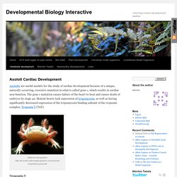
The gene c mutation causes failure of the heart to beat and causes death of embryos by stage 42. Mutant hearts lack expression of tropomyosin, as well as having significantly decreased expression of the tropomyosin binding subunit of the troponin complex, Troponin T (TnT). National Geographic Troponin T. Bulbus cordis. By the upgrowth of the ventricular septum the bulbus cordis is in great measure separated from the left ventricle, but remains an integral part of the right ventricle, of which it forms the infundibulum.
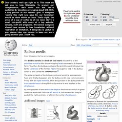
Additional images[edit] Head of chick embryo of about thirty-eight hours’ incubation, viewed from the ventral surface. X 26Diagram to illustrate the simple tubular condition of the heart.Heart of human embryo of about fourteen days.Human embryo about fifteen days old. Brain and heart represented from right side. Digestive tube and yolk sac in median section. References[edit] ON THE HEART OF THE ORANGE TUNICATE ECTEINASCIDIA TURBINATA HERDMAN. The Vertebrate Animal Heart: Unevolvable, whether Primitive or Complex. A Hearty Introduction: Before we get going, here are some exciting heart facts to start your day with: In case your momma never told you, hearts have 2 types of chambers: atria and ventricles.
Atria are where blood enters the heart and ventricles pump the blood out of the heart. Hearts take in oxygen poor blood, pump it through the pulmonary circuit (lungs/gills) where it gets oxygenated, and then they pump it out to the rest of the body. There are many many small veins (take blood to the heart) and arteries (carry it away) which connect the heart to the pulmonary circuit and keep it all running right.
Blood pressure has to be carefully balanced in all tubes so that flow pressure is maintained and blood keeps moving, but not so fast as to explode certain areas or capillaries. Given all this, there are 3 basic ways to make a heart found in animals: a 2 chambered heart, a 3 chambered heart, and a 4 chambered heart. Asymmetry in Embryonic development. At first glance, the left and right sides of our bodies are identical to one-another.
However, internally, some of our visceral organs are displaced to one side. The heart, for example, loops to the right side. This looping is consistent in the vertebrates and occurs very early in development. Rare individuals have the opposite symmetry, which is known as situs inversus. How is left-right asymmetry (often referred to as handedness) established during development?
In fact, a number of asymmetrically-expressed genes have been discovered, and we are on our way toward understanding how handedness develops. The demonstration of asymmetrical patterns of expression of these genes has made it possible to investigate the relationships among them so that regulatory interactions among them could be determined. Initially, Shh is expressed throughout Hensen's node. Asymmetric Shh expression induces a small domain of cells adjacent to the left side of the node to express nodal. Figure 1. Figure 2. Advanced - Molecular Development - Embryology. In attempts to determine the molecular mechanisms controlling heart development, scientists have focussed on early cardiomyocyte development including the cellular movement of cardiomyocyte progenitors and the signaling mechanisms that regulate cardiomyogenesis from the blastula to gastrula stages as well as the morphological changes that occur later in development such as looping and septation.
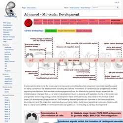
Some of the molecular and genetic factors regulating cardiac development have been previously described in this module in the aspect of development they pertain to. The following diagram provides an overview to the steps in cardiac development and the important associated genes, transcription factors and signalling molecules. Florida Museum of Natural History. What Does It Mean?

Genetics: the science of genes, heredity, and variation in living organisms. Systematics: the study of relationships among living things through time. Molecular Systematics: the analysis of hereditary molecular differences, mainly in DNA sequences, to gain information on an organism's evolutionary relationships. Research Highlights Plant Origins Angiosperms are amazingly diverse, with tens of thousands of living species. Image Galleries & Other Resources Cowrie Genetic Database Project This project has been established to rigorously examine the phylogenetic relationships within this diverse group of marine snails using a variety of DNA molecular markers.
Floral Genome Project. The conduction system. Advanced - Cardiac Conduction - Embryology. Endocardial Tube Cells Development in the Endocardium - LifeMap Discovery. Sinoatrial Node - Cellular Development, Function & Anatomy - LifeMap Discovery. Electrical conduction system of the heart. Principle of ECG formation.
