

Plotinus was a helluva cardshark. Beetle in a Box Five naked, blindfolded men get into a hottub.

The water represents the totality of facts, what we feel with our hands represents our picture of the world, and our penises... Didn't get the joke?
88 - Simplicity Itself: Plotinus on the One and Intellect. Plotinus. Biography[edit] Plotinus had an inherent distrust of materiality (an attitude common to Platonism), holding to the view that phenomena were a poor image or mimicry (mimesis) of something "higher and intelligible" [VI.I] which was the "truer part of genuine Being".

This distrust extended to the body, including his own; it is reported by Porphyry that at one point he refused to have his portrait painted, presumably for much the same reasons of dislike. Likewise Plotinus never discussed his ancestry, childhood, or his place or date of birth. From all accounts his personal and social life exhibited the highest moral and spiritual standards. Plotinus took up the study of philosophy at the age of twenty-seven, around the year 232, and travelled to Alexandria to study. Expedition to Persia and return to Rome[edit] Later life[edit] While in Rome Plotinus also gained the respect of the Emperor Gallienus and his wife Salonina. Major ideas[edit] Excitatory synapse. A diagram of a typical central nervous system synapse.

The spheres located in the upper neuron contain neurotransmitters that fuse with the presynaptic membrane and release neurotransmitters into the synaptic cleft. These neurotransmitters bind to receptors located on the postsynaptic membrane of the lower neuron, and, in the case of an excitatory synapse, may lead to a depolarization of the postsynaptic cell. An excitatory synapse is a synapse in which an action potential in a presynaptic neuron increases the probability of an action potential occurring in a postsynaptic cell. Neurons form networks through which nerve impulses travel, each neuron often making numerous connections with other cells.
These electrical signals may be excitatory or inhibitory, and, if the total of excitatory influences exceeds that of the inhibitory influences, the neuron will generate a new action potential at its axon hillock, thus transmitting the information to yet another cell.[1] Cytostasis. Cytostasis(cyto - cell, stasis - stoppage) is the inhibition of cell growth and multiplication.
Cytostatic refers to a cellular component or medicine that inhibits cell growth. Cytostasis is an important prerequisite for structured multicellular organisms. Electroporation. Electroporation, or electropermeabilization, is a significant increase in the electrical conductivity and permeability of the cell plasma membrane caused by an externally applied electrical field.
It is usually used in molecular biology as a way of introducing some substance into a cell, such as loading it with a molecular probe, a drug that can change the cell's function, or a piece of coding DNA.[1] Electroporation is a dynamic phenomenon that depends on the local transmembrane voltage at each point on the cell membrane. It is generally accepted that for a given pulse duration and shape, a specific transmembrane voltage threshold exists for the manifestation of the electroporation phenomenon (from 0.5 V to 1 V). This leads to the definition of an electric field magnitude threshold for electroporation (Eth). That is, only the cells within areas where E≧Eth are electroporated. Laboratory practice[edit] Poly ADP ribose polymerase. Members of PARP family[edit] The PARP family comprises 17 members (10 putative).
They have all very different structures and functions in the cell. Caspase. Caspases, or cysteine-aspartic proteases or cysteine-dependent aspartate-directed proteases are a family of cysteine proteases that play essential roles in apoptosis (programmed cell death), necrosis, and inflammation.[2] Types of caspase proteins[edit] As of November 2009[update], twelve caspases have been identified in humans.[3] There are two types of apoptotic caspases: initiator (apical) caspases and effector (executioner) caspases.

Initiator caspases (e.g., CASP2, CASP8, CASP9, and CASP10) cleave inactive pro-forms of effector caspases, thereby activating them. Effector caspases (e.g., CASP3, CASP6, CASP7) in turn cleave other protein substrates within the cell, to trigger the apoptotic process. The initiation of this cascade reaction is regulated by caspase inhibitors. Cytotoxic T cell. Antigen presentation stimulates T cells to become either "cytotoxic" CD8+ cells or "helper" CD4+ cells.
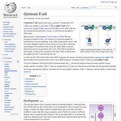
A cytotoxic T cell (also known as TC, cytotoxic T lymphocyte, CTL, T-killer cell, cytolytic T cell, CD8+ T-cells or killer T cell) is a T lymphocyte (a type of white blood cell) that kills cancer cells, cells that are infected (particularly with viruses), or cells that are damaged in other ways. Caspase 3. Caspase 3 is a caspase protein that interacts with caspase 8 and caspase 9.
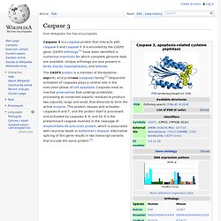
It is encoded by the CASP3 gene. CASP3 orthologs [1] have been identified in numerous mammals for which complete genome data are available. Unique orthologs are also present in birds, lizards, lissamphibians, and teleosts. Evidence for a membrane lipid defect in Alzheimer disease. Molecular Mechanisms Underlying Antiproliferative and Differentiating Responses of Hepatocarcinoma Cells to Subthermal Electric Stimulation. Capacitive Resistive Electric Transfer (CRET) therapy applies currents of 0.4–0.6 MHz to treatment of inflammatory and musculoskeletal injuries.
Previous studies have shown that intermittent exposure to CRET currents at subthermal doses exert cytotoxic or antiproliferative effects in human neuroblastoma or hepatocarcinoma cells, respectively. It has been proposed that such effects would be mediated by cell cycle arrest and by changes in the expression of cyclins and cyclin-dependent kinase inhibitors. The present work focuses on the study of the molecular mechanisms involved in CRET-induced cytostasis and investigates the possibility that the cellular response to the treatment extends to other phenomena, including induction of apoptosis and/or of changes in the differentiation stage of hepatocarcinoma cells.
Figures Editor: Irina V. Received: August 9, 2013; Accepted: November 15, 2013; Published: January 8, 2014 Copyright: © 2014 Hernández-Bule et al. Introduction.