

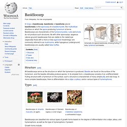
Basidiocarp. Schematic of a typical basidiocarp, showing fruiting body, hymenium and basidia Structure[edit] Types[edit] Basidiocarps of Ramaria rugosa, a coral fungus Basidiocarps are classified into various types of growth forms based on the degree of differentiation into a stipe, pileus, and hymenophore, as well as the type of hymenophore, if present.
Growth forms include: Basidium. Schematic showing a basidiomycete mushroom, gill structure, and spore-bearing basidia on the gill margins.
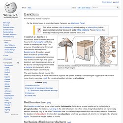
A basidium (pl., basidia) is a microscopic, spore-producing structure found on the hymenophore of fruiting bodies of basidiomycete fungi. The presence of basidia is one of the main characteristic features of the Basidiomycota. A basidium usually bears four sexual spores called basidiospores; occasionally the number may be two or even eight. In a typical basidium, each basidiospore is borne at the tip of a narrow prong or horn called a sterigma (pl. sterigmata), and is forcibly discharged upon maturity.
Basidium structure[edit] Mechanism of basidiospore discharge[edit] In most basidiomycetes, the basidiospores are ballistospores--they are forcibly discharged. Upon maturity of a basidiospore, sugars present in the cell wall begin to serve as condensation loci for water vapor in the air. Basidiospore. Basidiomycetes form sexual spores externally from a structure called a basidium.
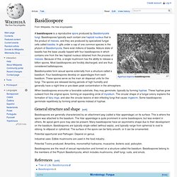
Four basidiospores develop on appendages from each basidium. These spores serve as the main air dispersal units for the fungi. The spores are released during periods of high humidity and generally have a night-time or pre-dawn peak concentration in the atmosphere. When basidiospores encounter a favorable substrate, they may germinate, typically by forming hyphae. These hyphae grow outward from the original spore, forming an expanding circle of mycelium. General structure and shape[edit] Basidiospores are generally characterized by an attachment peg (called a hilar appendage) on its surface.
Potential opportunist and Pathogen: Depend on genus. Industrial uses: Edible mushrooms are used in the food industry. Potential Toxins produced: Amanitins, monomethyl-hydrazine, muscarine, ibotenic acid, psilocybin. Conidium. Conidia on conidiophores Conidia, sometimes termed asexual chlamydospores, or chlamydoconida [1] are asexual,[2] non-motile spores of a fungus and are named after the Greek word for dust, skoni.
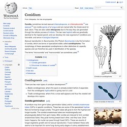
They are also called mitospores due to the way they are generated through the cellular process of mitosis. The two new haploid cells are genetically identical to the haploid parent, and can develop into new organisms if conditions are favorable, and serve in biological dispersal. Asexual reproduction in Ascomycetes (the Phylum Ascomycota) is by the formation of conidia, which are borne on specialized stalks called conidiophores. The morphology of these specialized conidiophores is often distinctive of a specific species and can therefore be used in identification of the species. The terms "microconidia" and "macroconidia" are sometimes used.[3] Sporangia & Zygosporangia. Home >> What fungi are >> How fungi reproduce >> Sexual reproduction >> Sporangia Among the true fungi only the Chytridiomycota and Zygomycota reproduce sexually by means of sporangia.
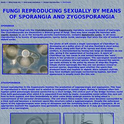
The Chytridiomycota are themselves a diverse group of fungi. They may have simple life histories with zygotic meiosis or, as in the mosquito parasite Coelomomyces, complex diplobiontic cycles. In all cases reproduction is by means of sporangiospores, spores borne inside sporangia that were the site of meiosis of diploid nuclei. The photo at left shows a single sporangium of Chytridium sp. developing on a pollen grain of red pine floating in pond water.
Sexual reproduction in the Zygomycota involves the production of zygosporangia and zygospores. The two pictures above show zygospores of Absidia spinosa, a common fungus in soil. Home >> What fungi are >> How fungi reproduce >> Sexual reproduction >> Sporangia. Bat-Killing Fungus Spreads West. Researchers have detected the fungus responsible for white-nose syndrome, which decimates bat populations, in Arkansas.
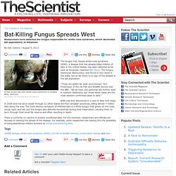
A little brown bat with white-nose syndrome in Greeley Mine, VermontWIKIMEDIA, US FISH AND WILFLIFE SERVICEThe fungus that causes white-nose syndrome (WNS), a disease that has already killed millions of bats in the United States, has been detected as far west as Arkansas, reported BBC News. The fungus, Geomyces destructans, was found in two caves in the state, but so far there is no sign of the disease in the bat population. “These are pretty far west occurrences,” Ann Froschauer of the US Fish and Wildlife Service told the BBC. “We do have one potential site further west in western Oklahoma, but these latest cases are the most western confirmed cases to date.”
WNS was first discovered in a cave in New York State in 2006 and has since swept through 22 other states and five Canadian provinces, killing almost 7 million bats along the way. Chytrid Fungus - The Frog Killer. Chytridiomycota. Life cycle & body plan[edit] Brief taxonomic history[edit] Habitats[edit] Ecological functions[edit] Batrachochytrium dendrobatidis[edit] The chytrid Batrachochytrium dendrobatidis is responsible for chytridiomycosis, a disease of amphibians.
Other parasites[edit] Chytrids mainly infect algae and other eukaryotic and prokaryotic microbes. Saprobes[edit] Arguably, the most important ecological function chytrids perform is decomposition.[9] These ubiquitous and cosmopolitan organisms are responsible for decomposition of refractory materials, such as pollen, cellulose, chitin, and keratin.[3][9] There are also chytrids that live and grow on pollen by attaching threadlike structures, the called rhizoids, onto the pollen grains.[21] This mostly occurs during asexual reproduction because the zoospores that become attached to the pollen continuously reproduce and form new chytrids that will attach to other pollen grains for nutrients.
Algae + Fungi = Symbiotic Lichen. High concentration of basidiocarps on moss-covered tree. Magic Mushrooms may have Cure for Depression.