

Mega Cisterna Magna Versus Arachnoid Cyst. Mega Cisterna Magna and Arachnoid are both types of cysts that occur in the central nervous system and contain cerebrospinal fluid, or CSF.

While they are very similar, it is important to make a correct diagnosis between the two in order to ensure proper planning and treatment and in order to properly diagnosis, a clear differentiation should be made. Both Mega Cisterna Magna and Arachnoid cysts account for less than one percent of all intracranial masses and can develop before or after birth. How They Are Defined A Mega cisterna magna refers to patients with enlarged CSF retrocerbellar cisterns in the posterior fossa, usually larger than 10mm in antenatal imaging. There will be normal cerebral morphology, also referred to as no abnormalities in the cerebellar. An arachnoid cyst is a sac filled with cerebrospinal fluid and located between the brain/spinal cord and the arachnoid membrane, one of three membranes that cover the brain and spinal cord.
Getting Properly Diagnosed. Bifocal intracranial tumors of nongerminomatous germ cell etiology: diagnostic and therapeutic implications. Kate's Brain surgery and recovery help by Kate N Quique Rivera. I am a 33 year old, mother of 4 bioligical children, and 2 step children.

The majority of my time and life have revolved around my children and have always enjoyed being active... up until 2008, I started having horrible migraines, blurred vision, constant state of fog and confusion, vertigo, balance and coordination issues, ringing in my ears, constant headache and pressure behind my eyes and in my ears, light and sound sensitivity, visual disturbances (double vision, blurry vision and problems focusing my eyes), and constant fatigue, which progressivly worsened. The doctors kept diagnosing me with migraines. I tried medication after medication, none of which helped with the symptoms. Surgery for pineal cysts?
I have never in my career, 43 years, found it necessary to operate on a pineal cyst.

I have operated on over 100 pineal tumors in children none of them being pineal cysts. The incidence of asymptomatic pineal cysts at autopsy is 10%.Of course it possible that the occasional pineal cyst may cause acqueductal occlusion but this is very rare. The aqueduct often looks distorted but rarely is occluded. if the cyst is truly causing progressive hydrocephalus then endoscopic fenestration is the simplest and safest approach through the dilated ventricles.I wonder if anyone has a case of un-operated pineal cyst where any untoward clinical outcome has occurred.
Pineal Region Tumors – Neurosurgical Review - ScopeMed.org - Online Journal Management System. Pubmed Style Radovanovic I, Dizdarevic K, Tribolet Nd, Masic T, Muminagic S.

Pineal Region Tumors – Neurosurgical Review . Med Arh. 2009; 63(3): 171-173. Leptomeningeal Carcinomatosis. Signs and symptoms Meningeal symptoms are the first manifestations in some patients (pain and seizures are the most common presenting complaints) and can include the following: Headaches (usually associated with nausea, vomiting, light-headedness)Gait difficulties from weakness or ataxiaMemory problemsIncontinenceSensory abnormalities CNS symptoms are divided into the following 3 anatomic groups:
Internet Scientific Publications. Hormones.gr. Case report Metastatic bronchial neuroendocrine tumor to the pineal gland: A unique manifestation of a rare disease Simona Grozinsky-Glasberg1,3, Susana Fichman2, Ilan Shimon1,3 1Institute of Endocrinology and 2Institute of Pathology, Beilinson Hospital, Rabin Medical Center; and 3Sackler Faculty of Medicine, Tel Aviv University, Israel Abstract.
Press release: Contrast Agent Linked with Brain Abnormalities on MRI. CHICAGO – For the first time, researchers have confirmed an association between a common magnetic resonance imaging (MRI) contrast agent and abnormalities on brain MRI, according to a new study published online in the journal Radiology.
The new study raises the possibility that a toxic component of the contrast agent may remain in the body long after administration. Brain MRI exams are often performed with a gadolinium-based contrast medium (Gd-CM). Gadolinium's paramagnetic properties make it useful for MRI, but the toxicity of the gadolinium ion means it must be chemically bonded with non-metal ions so that it can be carried through the kidneys and out of the body before the ion is released in tissue. Gd-CM is considered safe in patients with normal kidney function. To learn more, the researchers compared unenhanced T1-weighted MR images (T1WI) of 19 patients who had undergone six or more contrast-enhanced brain scans with 16 patients who had received six or fewer unenhanced scans.
Pineal Brain Tumor: Patient's Perspective · Healthy Living articles. Welcome to the Center for Neuroacoustic Research! Dr.
Jeffrey D. Thompson, D.C., B.F.A. Pineal Tumor - Treatment UCLA. General Information.
Papillary tumors of the pineal region. Papillary tumors of the pineal region (PTPR) were first described by A.
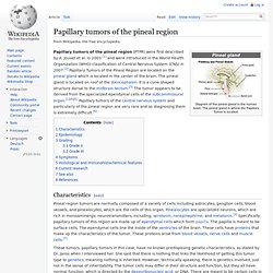
Jouvet et al. in 2003 [1] and were introduced in the World Health Organization (WHO) classification of Central Nervous System (CNS) in 2007.[2] Papillary Tumors of the Pineal Region are located on the pineal gland which is located in the center of the brain. The pineal gland is located on roof of the diencephalon. It is a cone shaped structure dorsal to the midbrain tectum.[3] The tumor appears to be derived from the specialized ependymal cells of the subcommissural organ.[1][4][5] Papillary tumors of the central nervous system and particularly of the pineal region are very rare and so diagnosing them is extremely difficult.[6] Characteristics[edit] Pineal Region Tumors - New York Presbyterian Hospital. Pineal Region Tumors. Search Results.
Magnetic resonance imaging of pineal region tumours. Human pineal physiology and functional significance of melatonin. Macchi MM, Bruce JN. Neuroradiology Cases: Meckel's Cave Lipoma MRI. A young male patient with right sided trigeminal neuralgia clinically. MRI study of Brain (Axial FLAIR, T2w, T1w, T2*GRE and STIR) This MRI study of Brain shows: A lobulated well circumscribed T1 hyper intense tissue at right CP angle extending in middle cranial fossa in right para sellar region through Meckel’s cave, along the course of Trigeminal nerve giving it a so called dumbbell shape.
Signals are isointense to subcutaneous fat on T1w and T2w images with complete signal suppression on STIR corresponds to 'fat', it’s a lipoma. Low signals on T2w *GRE is due to an associated calcification which is known. Neuroradiology Cases: Wernicke's Encephalopathy MRI. A 70 yo female, known case of ca cervix with metastasis. Pt relative complaints of her neglect towards food. Vomits out anything she eat.
Now brought unconscious. Axial FLAIR shows an abnormal T2 hyperintensity involving mid brain in the region of peri aqueductal grey matter and hypothalamus. Imaging findings support changes of Wernicke's Encephalopathy. In WE, CT Brain is often normal. 3908.full. Brain-derived neurotrophic factor. Brain-derived neurotrophic factor, also known as BDNF, is a secreted protein[2] that, in humans, is encoded by the BDNF gene.[3][4] BDNF is a member of the "neurotrophin" family of growth factors, which are related to the canonical "nerve growth factor", NGF. Neurotrophic factors are found in the brain and the periphery.
Function[edit] THE BRAIN FROM TOP TO BOTTOM. Melatonin, the hormone secreted by the pineal gland, was not discovered until the late 1950s. Its role in regulating biological rhythms was then uncovered only gradually, making the pineal gland the last of the endocrine glands to have its function identified. HEAD%20IMAGING%20GUIDELINES%202011.
DBH - dopamine beta-hydroxylase (dopamine beta-monooxygenase) Reviewed September 2008. Genetically determined dopamine availability predicts disposition for depression. Brain and Nerves. Brain & Nervous System Disorders Board Index: mega cisterna magna. ... So, what did he say? Datasheet_dna_methylation_analysis. Epigenomics Fact Sheet. Epigenomics What is the epigenome? A genome is the complete set of deoxyribonucleic acid, or DNA, in a cell. DNA carries the instructions for building all of the proteins that make each living creature unique. Derived from the Greek, epigenome means "above" the genome. Manic Episode Associated with Mega Cisterna Magna.
Levin_ch07_p193-207. Pineal cyst. Pineal cysts are common, usually asymptomatic, and typically found incidentally. Their importance is mainly in the fact that they cannot be distinguished from cystic tumours, especially when large or when atypical features are present. As such, many patients undergo prolonged followup for these lesions, presumably with associated anxiety. Pineal Brain Tumor: Patient's Perspective · Healthy Living articles. Pineal Gland Cysts. Unfortunately, there really is no treatment. Many people suffering from symptoms are treated flippantly by their doctors, neurologists, and neurosurgeons.
Gland Disorders Forum - Pineal Cyst. I started having headaches, daily and neck/ear/jaw pain back in May. Johns Hopkins Medicine Health Library. What is a brain tumor? PHYSIOLOGY OF THE PINEAL GLAND. Neurosurgical Perspective. The Pineal Gland and Pineal Tumours - Chapter 15 : NEUROENDOCRINOLOGY, HYPOTHALAMUS, AND PITUITARY.